
Understanding Radiation-Free Bone Density Testing Technology
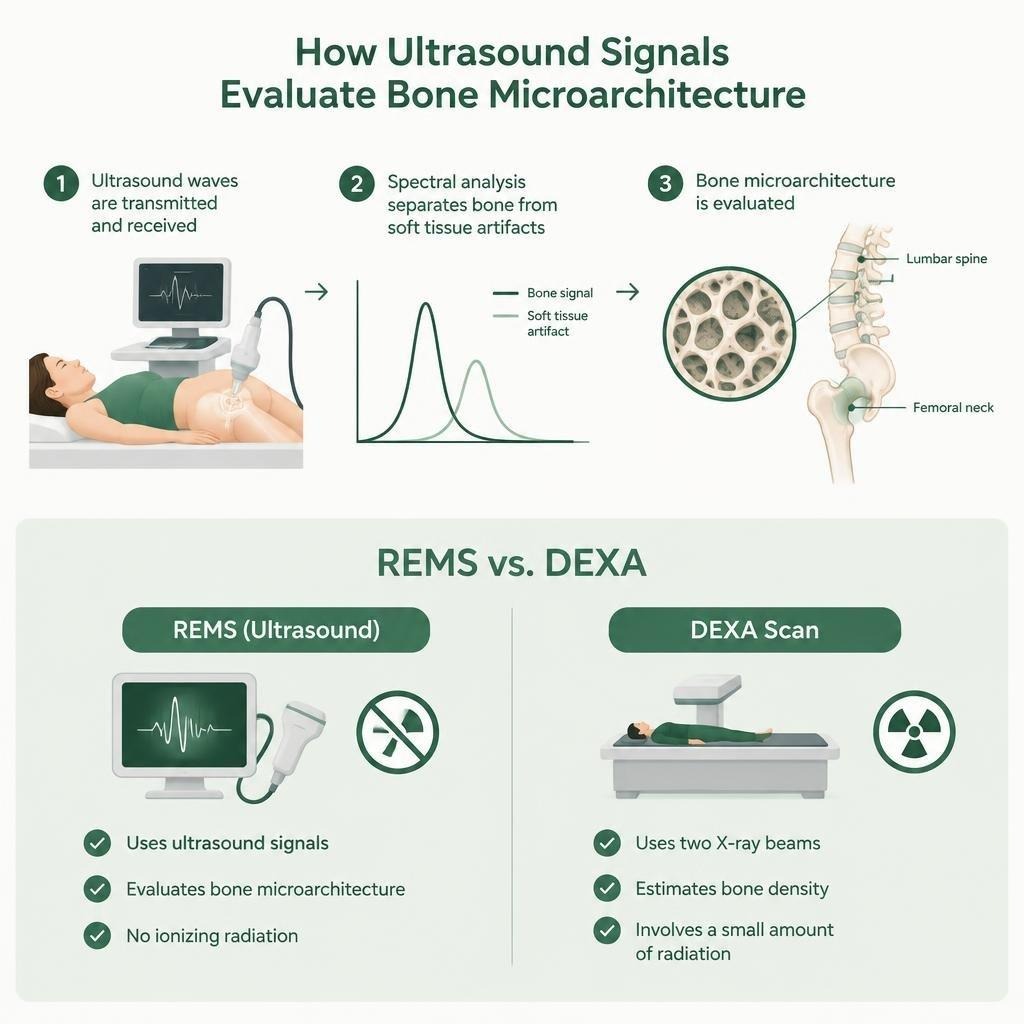
At the heart of this innovation is Radiofrequency Echographic Multi Spectrometry (REMS). Unlike traditional imaging that relies on ionizing radiation, REMS utilizes ultrasound signals to analyze the internal structure of the bone. By transmitting and receiving ultrasound waves, the technology evaluates the bone microarchitecture at the lumbar spine and femoral neck—the two most critical sites for assessing fracture risk.
The diagnostic accuracy of this technology stems from its ability to perform a spectral analysis of the raw ultrasound signals. This process allows the system to distinguish between bone tissue and surrounding soft tissue artifacts, which can sometimes interfere with the precision of other imaging methods. Because it does not rely on radiation, it is particularly well-suited for patients who require frequent monitoring, such as those undergoing long-term treatment for osteopenia or osteoporosis, or individuals who are simply looking to establish a baseline for their skeletal health without cumulative radiation exposure.
How Radiation-Free Bone Density Testing Compares to Traditional DEXA Scans
For decades, the Dual-Energy X-ray Absorptiometry (DEXA) scan has been the clinical “gold standard” for measuring bone mineral density. However, it is important to understand the fundamental differences between this legacy technology and modern ultrasound-based approaches.
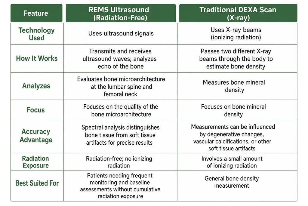
DEXA works by passing two different X-ray beams through the body to estimate bone density. While highly effective, it does involve a small amount of ionizing radiation. Furthermore, DEXA measurements can occasionally be influenced by factors like degenerative changes, vascular calcifications, or other soft tissue artifacts that might lead to an overestimation of bone density.
In contrast, ultrasound-based testing focuses on the quality of the bone microarchitecture. By analyzing the “echo” of the bone, the technology provides a comprehensive look at the skeletal structure.
Clinical Applications and Patient Eligibility
Who stands to benefit most from this technology? The primary candidates are individuals focused on long-term bone wellness. This includes postmenopausal women and men over the age of 50 who are monitoring for age-related bone loss.
Beyond general screening, this technology is valuable for those managing secondary osteoporosis, which can be caused by certain medications or underlying metabolic bone disorders. Because the scan is non-invasive and radiation-free, it is an excellent tool for baseline assessments. By starting with a clear, accurate scan, patients can work with their healthcare providers to track bone health trends over time, ensuring that any necessary interventions are identified as early as possible.
Key Diagnostic Advantages and Procedure Details
One of the most significant advantages of this technology is the patient experience. Because the procedure is non-invasive and requires no ionizing radiation, it is comfortable and straightforward. During the scan, a technician applies a standard acoustic coupling gel to the skin, similar to what you might experience during a routine pregnancy ultrasound.
What to Expect During Radiation-Free Bone Density Testing
When you arrive for your appointment, the process typically takes between 15 to 30 minutes. You will be asked to lie comfortably on an examination table while the technician positions the ultrasound probe over the target areas—usually the lower back and hip.
There is no need for special preparation, such as fasting or changing into a medical gown, provided you are wearing comfortable clothing that allows easy access to the scan sites. The system provides real-time feedback, allowing the technician to ensure the data capture is accurate. Once the scan is complete, the data is processed, and a report is generated that highlights your bone health metrics, which you can then discuss with your physician.
Monitoring Bone Health and Scan Frequency Over Time
Consistency is key when tracking bone health. Because this method is safe and radiation-free, it allows for more frequent longitudinal tracking if your doctor recommends it. Rather than waiting years between scans, you and your healthcare provider can monitor how your bone quality metrics change in response to lifestyle adjustments, nutritional changes, or therapeutic interventions. This ability to track microarchitecture scoring over time provides a more nuanced view of your skeletal health than a simple density number alone.
How to Find and Choose Certified Scanning Facilities
Finding a provider that offers this technology is becoming easier as more clinics adopt non-invasive diagnostic tools. When searching for rems scan locations near me, it is important to look for facilities that prioritize patient care and clear communication of results.
When evaluating a facility, consider the following criteria:
- Technologist Training: Ensure the staff is specifically trained in the operation of the ultrasound-based bone density technology.
- Equipment Calibration: Ask if the equipment is regularly serviced and calibrated to ensure the highest diagnostic precision.
- Report Delivery: A good facility will provide a clear, comprehensive report that is easy for both you and your primary care physician to understand.
- Follow-up Care: Choose a center that offers a consultation or a clear path to discuss your results with a qualified professional.
While some specialized clinics offer these services, you can also explore broader resources to find rems scan locations near me that might be closer to your home or office. Don’t hesitate to call ahead and ask specific questions about their experience with this technology.
Frequently Asked Questions About Bone Density Screening
How accurate are ultrasound-based bone scans compared to traditional x-ray methods?
Clinical validation studies have shown that ultrasound-based bone density testing offers high diagnostic precision. By using echographic spectrometry, the technology provides a reliable assessment of bone mineral density. While it differs from the X-ray approach, it is designed to provide a highly accurate estimation of bone strength and fracture risk.
Does medical insurance cover non-radiation bone density scans?
Coverage varies significantly depending on your specific insurance provider and policy. Some diagnostic scans are covered under standard imaging benefits, while others may be classified differently. It is always a good idea to check if your facility accepts your insurance or if you can use funds from a Health Savings Account (HSA) or Flexible Spending Account (FSA) to cover the cost. Many clinics also offer competitive cash-pay rates for those without insurance coverage.
What questions should patients ask a scan provider prior to booking?
Before scheduling, consider asking:
- “How long have you been performing this specific type of bone density scan?”
- “Will I receive a copy of the results to take to my primary doctor?”
- “Is there a qualified professional available to help me interpret the findings?”
- “What is the total cost, and do you offer payment plans or insurance filing assistance?”
Conclusion
Taking charge of your bone health is a proactive step toward maintaining your independence and quality of life. By choosing modern, radiation-free diagnostic options, you can monitor your skeletal integrity with confidence and clarity. Whether you are seeking a baseline measurement or tracking the progress of a bone health program, the ability to access accurate, non-invasive imaging is a powerful tool in your wellness arsenal. As you research the best options for your needs, the most important factor is consistent monitoring and an open dialogue with your healthcare team.